Tuesday, April 27, 2010

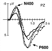
Training of Executive Control Functions: Negative Transfer and Far Transfer


The Cognitive Neuroscience Society 2010 Annual Meeting was held last week in Montreal, Québec. Unfortunately, many European registrants were unable to attend because of the Eyjafjallajokull volcano. The meeting website has links to the PDFs for 67 of these "Volcano Posters".

One of those unable to attend was Dr. Jonas Persson of Stockholm University. He was scheduled to speak in the final symposium of the conference, which was on control of executive control, or who controls the "controller" in the brain (without resorting to a homunculus or an infinite regress of Mini-Me's). His co-author, Dr. Patricia Reuter-Lorenz of the University of Michigan, gave an interesting presentation about their work on the ups and downs of cognitive training: gains that transfer to other tasks across sessions and fatigue that transfers to other tasks within a session. The abstract is reprinted below.

---------------
Symposium Session 5
Tuesday, April 20, 1:00 - 3:00 pm, Westmount et al Ballroom

What Controls Executive Control? The Influence of 'Control Context'

Chair: Amishi Jha, University of Pennsylvania

. . .

Talk 3: Training and Depletion of Executive Functions: The Case of Interference Control

Jonas Persson1, Patricia Reuter-Lorenz2; 1Stockholm University, 2University of Michigan

Brain imaging reveals overlapping sites of prefrontal activation for different cognitive tasks suggesting they may share core executive processes. We tested this hypothesis by measuring behavioral interactions between memory tasks presumed to require interference control - a putative executive process that mediates selection from competing representations. Behavioral data show that different training regimens produce either negative or positive transfer from working memory to semantic and episodic memory task performance. We show that eight days of training on high interference versions of three different working memory tasks increased the efficiency of interference control on the training tasks and on untrained tasks in new memory domains. In contrast we have also demonstrated negative transfer and process-specific “fatigue” effects indicating that control efficiency in a second task is diminished by high control demands in a prior task immediately preceding it in time. This suggests that interference control is a finite resource that can be temporarily depleted. Functional magnetic resonance imaging (fMRI) was used to elucidate the mechanisms associated with decreasing efficiency or resource depletion of the interference control process. Along with reduced performance, fMRI indicates negative transfer is associated with reduced process-specific activation, and increased homologous activation that may be compensatory. In sum, this suggests that interference control is an executive function that is both resource limited and plastic making it possible for training to alter its efficiency.
---------------

These findings became extremely relevant (albeit ignored) in light of the ultra-high impact paper by Adrian Owen et al. (2010) published in BBC/Nature on April 20, informing us there was "No gain from brain training" in a massive group of 11,430 volunteers. The research volunteers participated in online training exercises that tapped (1) reasoning, planning, problem solving; (2) short-term memory, attention, visuospatial processing, mathematics; or (3) ability to answer obscure questions using the internet.


Numerous other outlets have already summarized these results, so I won't attempt to do so here. The main issue was whether the gains obtained through simple practice effects transferred to tasks that weren't in the training set. The short answer is no.

Brain training doesn't boost brain power, work suggests (BBC)

Brain-training games don't work (Guardian)

Brain training games don't work (BPS Research Digest)

Brain-training games get a D at brain-training tests (Not Exactly Rocket Science)

Brain Test Britain - Results in-depth (BBC - Lab UK)

I am not a proponent of brain training games that make unsound claims lacking credible scientific evidence to back them up. But in light of the spectacularly negative findings of Owen et al., the question arises of whether their training regimen (10 min 3 times a week for 6 weeks) was adequate to produce significant effects. Predictably, those with a stake in the matter said no, the training was neither sufficient in duration nor applicable to other commercially available products.

BBC “Brain Training” Experiment: the Good, the Bad, the Ugly

Posit Science Disputes Results of the BBC Brain Training Study

A final issue concerns the population under study, a presumably "normal" group of 18-60 year olds without cognitive impairments or cognitive decline. Would the same training exercises help an older population at risk for dementia? On the other hand, seniors might be especially vulnerable to persuasive false claims from unscrupulous brain training vendors. Thus it's important to have scientifically valid and accessible research on whether cognitive training might help an aging population (Lustig et al., 2009). Coincidentally, the NIH is currently trying to reach a consensus on:
NIH State-of-the-Science Conference
Preventing Alzheimer’s Disease and Cognitive Decline

April 26–28, 2010
Bethesda, Maryland


Live webcast, also information on archived video and publicly available consensus statement.
Let us now return to the studies of Persson and Reuter Lorenz (2008). This work examined whether the training of a specific cognitive process (overcoming interference from competing representations in memory) in one task will improve the ability to overcome interference in a different task. For example, a set of four letters would be presented in a working memory task, followed by a delay and then a probe letter, which requires a yes/no response (was this letter a part of the set you just studied?). The interference arises when participants must reject a probe letter that was a member of the memory set on the preceding trial, but is not a member of the set on the current trial ("Recent Negative", illustrated below).


Figure 1 (D'Esposito et al., 1999). Target items were presented for 950 msec followed by a 7,050-msec delay period. This interval was followed by a probe letter for 1,500 msec that either was recently presented (Recent trial) or was not recently presented (Nonrecent trial). Note that, in the Recent Negative trial, the probe letter “P” is not in the target set of that trial but that it is in the target set of the two previous trials.

In the current experiment (Persson & Reuter-Lorenz, 2008), 48 young control participants were randomly assigned to one of three groups:
  1. Interference Training (working memory requirements and interference)
  2. Control 1 (working memory requirements only)
  3. Control 2 (minimal memory requirements and no interference)
Training consisted of eight 40 min sessions over a two week period. Benchmark tests were administered on the days before and after the training program to evaluate interference resolution performance on non-trained tasks of working memory (item recognition with concrete nouns), semantic memory (verb generation), and episodic memory (paired associates). The experimental design is shown below.


Fig. 1 (Persson & Reuter-Lorenz, 2008). Schematic depiction of the basic experimental design. The illustration shows the tasks performed by each subject group during each phase of the experiment: pretraining (Session 1), training (Sessions 2–9), and posttraining (Session 10). All groups performed the same three transfer tasks in the pretraining and posttraining sessions. During training, the interference-training group performed tasks involving working memory (WM) and interference; Control Group 1 performed noninterference versions of the same tasks, and Control Group 2 performed similar tasks that required little WM and involved no interference.

Results indicated that Interference Training reduced reaction time (RT) measures of interference in each of the three transfer tasks (left bars), but control training did not (right bars).

Fig. 3. (Persson & Reuter-Lorenz, 2008). Mean interference-resolution scores (response time in the high-interference condition minus response time in the low-interference condition) on the transfer tasks as a function of group (interference-training group vs. control groups) and time (pretraining vs. posttraining). Results are shown separately for the (a) verb-generation task (semantic memory), (b) item-recognition task (working memory), and (c) paired-associates task (episodic memory). Error bars show standard errors of the means.

A previous study demonstrated the opposite sort of process-specific interactions, in the form of within-session fatigue effects (i.e., negative transfer) across a subset of these tasks (Persson et al., 2007):
The idea that tasks with overlapping neural representations may involve similar executive components was ... critical to our approach. Of particular interest were tasks requiring resolution of interference among competing representations. Within a single experimental session intensive training reduced the ability to resolve interference on a transfer task if the training task placed high demands on interference resolution. Negative transfer was absent when interference resolution was minimally required by the task, or when the training and transfer tasks did not rely on overlapping neural representations.
Based on these results, the authors concluded that targeted training -- which identifies and trains a specific cognitive process that is implemented by a known network in the brain -- can produce improvements (across sessions) that transfer to tasks other than those in the training set. Conversely, fatigue effects (within session) can result in negative transfer to related tasks. In the case of interference resolution for items held in memory (or "proactive interference"), the neuroimaging literature suggests that a particular area in the inferior frontal gyrus of the left frontal lobe is a key region for overcoming interference among competing memory representations in various types of tests (Jonides & Nee, 2006).

What does all this mean for the brain training biz and for the BBC/Nature study? Adequate training time and a brain-based approach to target highly specific cognitive processes and tasks will yield the best results.


ADDENDUM (April 10, 2011): The 2008 Psychological Science paper by Persson and Reuter-Lorenz has been retracted:
This article has been retracted by both of the authors. During further extension of this work, it was discovered that a mistake had been made in the programming of the working memory training tasks used in this study. This error resulted in the presentation of repeated trial sequences, which were then practiced repeatedly over the 8-day training period. This practice likely resulted in learning of the sequences, rather than training of interference control as we had originally inferred. The first author takes responsibility for this error, and both authors regret the publication of invalid results. In a separate replication (N = 16) of the working memory training condition that used randomized, novel sequences for each of the 8 training days, we were not able to reproduce the main findings of the original report...

References

Jonides J, Nee DE. (2006). Brain mechanisms of proactive interference in working memory. Neuroscience 139:181-93.

Lustig C, Shah P, Seidler R, Reuter-Lorenz PA (2009). Aging, training, and the brain: a review and future directions. Neuropsychol Rev 19:504–522.

Owen AM, Hampshire A, Grahn JA, Stenton R, Dajani S, Burns AS, Howard RJ, Ballard CG. (2010). Putting brain training to the test. Nature Apr 20. [Epub ahead of print]. PDF

Persson, J., & Reuter-Lorenz, P. (2008). Gaining Control: Training Executive Function and Far Transfer of the Ability to Resolve Interference. Psychological Science 19: 881-888. DOI: 10.1111/j.1467-9280.2008.02172.x

PERSSON, J., WELSH, K., JONIDES, J., & REUTER-LORENZ, P. (2007). Cognitive fatigue of executive processes: Interaction between interference resolution tasks. Neuropsychologia 45: 1571-1579. DOI: 10.1016/j.neuropsychologia.2006.12.007

Subscribe to Post Comments [Atom]

Tuesday, April 13, 2010

Mirror Neurons Join Marilyn Monroe Neurons and Halle Berry Neurons in the Human Hippocampus


Move over, Marilyn Monroe neurons and Halle Berry neurons... The cellular media darlings of action observation and action execution would like to join you in the human hippocampus and surrounding medial temporal lobe (MTL) areas critical for memory.

"What?" you say. "Direct evidence for the existence of mirror neurons has been obtained from single cell recordings in monkeys in specific brain regions. These include the ventral premotor cortex (area F5) and the inferior parietal lobule (Rizzolatti & Sinigaglia, 2010), not the hippocampus!"


Figure 1 (Rizzolatti & Sinigaglia, 2010): The parieto-frontal mirror network. Lateral view of the macaque brain. The coloured areas represent the areas of the parieto-frontal circuit containing mirror neurons: the ventral premotor cortex (area F5), area PFG (located between parietal areas PF and PG) and the anterior intraparietal area (AIP)... The parieto-frontal circuit receives high-order visual information from areas located inside the superior temporal sulcus (STS) and the inferior temporal lobe (IT). Neither of these temporal regions has motor properties. The parieto-frontal circuit is under control of the frontal lobe (area F6 or pre-supplementary motor area and the ventral prefrontal cortex (VPF)). The inset provides an enlarged view of area F5. IAS, inferior limb of the arcuate sulcus; LIP, lateral intraparietal area; VIP, ventral intraparietal area.

Yet we've been led to believe that a new study published in Current Biology (Mukamel et al., 2010) has recorded from mirror neurons in the human brain for the first time. Last Friday, BPS Research Digest asked:
Is this the first ever direct evidence for human mirror neurons?

...Although recordings from single cells in the brains of monkeys have identified 'mirror' neurons that respond both to the execution of a movement and the observation of another agent performing that same movement, the existence of such cells in humans has, up until now, been inferred only from indirect evidence, particularly brain imaging. Now Roy Mukamel and colleagues have provided what appears to be the first ever direct evidence, using implanted electrode recordings of single cells, for the existence of mirror neurons in humans.
The short answer to this question is "no" (unless you want to dilute the meaning of "mirror neurons" beyond recognition). To briefly summarize, the participants in the study were 21 patients with pharmacologically intractable epilepsy. Depth electrodes were implanted into their brains to monitor for seizures, in advance of a possible surgical intervention to remove the epileptic focus. The electrode locations were constrained by clinical considerations and included regions in the medial frontal cortex (supplementary motor area, anterior cingulate cortex) and the medial temporal lobe (amygdala, hippocampus, parahippocampal gyrus, entorhinal cortex). The ventral premotor cortex and inferior parietal lobe were not targeted.

The experimental protocol consisted of 3 tasks: Grasp, Facial expressions and Control.
During Grasp, subjects were presented with video clips of a hand grasping a mug and with the words ‘Finger’ or ‘Hand’. They were instructed to grasp a mug with precision grip or whole hand prehension when the words ‘Finger’ or ‘Hand’, respectively, were presented and to simply observe when the video clips were played. During Facial expressions, subjects were presented with a picture of a smiling or a frowning face and with the word ‘Smile’ or ‘Frown’. They were instructed to perform the corresponding action when the words were presented and to simply observe when the pictures were presented... In the Control task, subjects were presented with the words used as cues in the Grasp and Facial expression parts of the experiment and were instructed to covertly read the words and refrain from making hand movements or facial gestures.
Results are depicted in Table 1 below (click on image for a larger view).

In each brain region, only a minority of cells responded with an increased (or decreased) firing rate during both observation and execution of the same action. Percentages of these Observation/Execution neurons ranged from a low of 2% in dorsal anterior cingulate cortex to 11% in hippocampus, 12% in parahippocampal gyrus, and 14% in supplementary motor area. The MTL regions also contain neurons that respond during the spontaneous recall of episodic memories (Gelbard-Sagiv et al., 2008). So how can you tell if a neuronal response in the current experiment is related to memory recall or to action observation/execution? You can't, but that doesn't matter!
The action observation/execution matching neurons in the medial temporal lobe may match the sight of actions of others with the memory of those same actions performed by the observer. Thus during action-execution, a memory of the executed action is formed, and during action-observation this memory trace is reactivated. This interpretation is in line with the hypothesis of multiple mirroring mechanisms in the primate brain, a hypothesis that can easily account for the presence of mirroring cells in many cortical areas.
"Now wait a minute," said Professor Patricia Churchland [as paraphrased by Prof. Greg Hickok in Talking Brains]. "If mirror neurons are all over the brain then don't they lose their explanatory power?"

Good point.

Another issue is whether a given single- or multi-unit recording responded by increasing or decreasing its firing rate relative to the passive condition. It could be either, or both:
Among the 68 action observation/execution matching cells [out of 1177 total cells recorded], 33 increased their firing rate during both observation and execution of a particular action. In contrast, 21 other neurons decreased their firing rate during both conditions. These types of responses have been previously reported in monkeys and birds. Furthermore, 14 neurons increased their firing rate during one condition and decreased it during the other.
Really? Neurons can show inhibitory responses to observation and execution, or mismatched responses, and still be considered "mirror neurons"?

Also notable is that the paper did not refer to previous results from the same lab on Marilyn Monroe neurons (Quian Quiroga et al., 2009) and Halle Berry neurons (Quian Quiroga et al., 2005).1 The new "mirroring cells" are apparently intermixed with individual neurons that show hyperspecific responses to pictures of celebrities (taken from various angles, in and out of character) and even to their printed names and voices. In each brain region, about 10% of the cells were responsive to stimulation of any sort. Of these minority neurons, 0% in parahippocampal cortex, 14% in amygdala, 35% in entorhinal cortex, and 38% in hippocampus showed "triple invariance" to presentation as pictures, sound, and text (Quian Quiroga et al., 2009).

The hyperspecific neuronal responses included a Jennifer Aniston+Brad Pitt cell (not Aniston alone), a Pamela Anderson cell that responded to a caricature of her and to her printed name, and a Kobe Bryant cell. A specific double dissociation was reported between a Halle Berry neuron and a Mother Teresa neuron (i.e, one cell showed a response to Halle Berry but not Mother Teresa, and the other cell showed a response to Mother Teresa but not Halle Berry).

Below is one of my favorites, the rare Robert Plant neuron that responds to images, sounds, and text depicting the former Led Zeppelin singer. I wonder what would happen if he had a closed shirt and shorn hair in some of the pictures?


Figure S17 (Quian Quiroga et al., 2009). A single unit in the entorhinal cortex selectively activated by pictures, sound and text presentations of Robert Plant, singer of the band ‘Led Zeppelin’, which was known to the patient.

But what about hybrid Halle Berry mirror neurons? How does one integrate the results from all these studies? Who's to say that you couldn't find a Jennifer Aniston action observation/action imitation neuron if you looked hard enough? Would it still be considered a "mirroring neuron" if Lisa Kudrow did not elicit the same response? What if all the cast members of Friends could evoke the observation/imitation response, but not the cast members of Seinfeld?

In conclusion, Mukamel et al., (2010) have this to say about their "mirror neuron" results:
The functional significance of the mirror mechanism most likely varies according to the location of mirror neurons in different brain areas. For example, the mirror mechanism in the insula might underlie the capacity to understand a specific emotion (disgust) in others, whereas the mirror mechanism in the parietofrontal circuit may help understanding the goal of observed motor acts and the intentions behind them. Here we show cellular mirroring mechanisms in areas relevant to movement initiation and sequencing (SMA) and to memory (medial temporal lobe). Whereas these hypotheses have yet to be tested more carefully, these results demonstrate the presence of mirror mechanisms in humans at the single neuron level and in areas functionally different from the ones previously described in the literature.
So are mirror neurons everywhere? Have they lost their explanatory power?


Footnote

1 One of the Fried lab papers was cited in passing but only to say how the new "mirror neuron" results were not due to representational invariance of grasping or smiling, because cell firing rates did not change in the passive control condition. However, the lack of a significant response in the control condition was required before including a cell(s) in the action execution bin. I can imagine that quite a few neurons responded in a condition where subjects were told, "covertly read the words and refrain from making hand movements or facial gestures" -- especially in the SMA and pre-SMA, which are involved not only in motor planning but in motor inhibition as well.

References

Gelbard-Sagiv H, Mukamel R, Harel M, Malach R, Fried I. (2008). Internally generated reactivation of single neurons in human hippocampus during free recall. Science 322:96-101.

Mukamel, R., Ekstrom, A., Kaplan, J., Iacoboni, M., & Fried, I. (2010). Single-Neuron Responses in Humans during Execution and Observation of Actions. Current Biology DOI: 10.1016/j.cub.2010.02.045


Quian Quiroga, R., Kraskov, A., Koch, C., & Fried, I. (2009). Explicit Encoding of Multimodal Percepts by Single Neurons in the Human Brain. Current Biology, 19 (15), 1308-1313 DOI: 10.1016/j.cub.2009.06.060

Quiroga, R., Reddy, L., Kreiman, G., Koch, C., & Fried, I. (2005). Invariant visual representation by single neurons in the human brain. Nature, 435 (7045), 1102-1107 DOI: 10.1038/nature03687

Rizzolatti G, Sinigaglia C. (2010). The functional role of the parieto-frontal mirror circuit: interpretations and misinterpretations. Nat Rev Neurosci. 11:264-74.

Subscribe to Post Comments [Atom]

Sunday, April 11, 2010

"Sleeping Beauty Paraphilia" and Body Image Disturbance After Brain Injury

Brain injuries caused by strokes, tumors or head trauma can, on occasion, result in Unusual Changes in Sexuality, as discussed in an earlier blog post. A new case report by Bianchi-Demicheli et al. (2010) describes a unique paraphilia1 in a married 34 year old man. The authors called it Sleeping Beauty paraphilia:
This [man] felt sexually aroused from seeing sleeping women as well as from taking care of their hands and nails while they were asleep.
The patient came to the attention of the authors when he was brought to the emergency psychiatric unit after assaulting his wife with pepper spray while wearing a latex mask.2 More details of the case are as follows:
His marriage had been in crisis for several years because over the time the patient developed a particular and progressive sexual deviant behaviour. He felt sexually aroused from seeing sleeping women as well as from taking care of their hands and nails while they were asleep, beginning mostly with the right hand.

In the first time of his marriage he could control these fantasies, but over the years he lost the control of his sexual urges and he must irresistibly act his deviant behaviour. In order to realize his uncontrollable impulse, he was used to provide his wife sleeping pills to satisfy his paraphilia. In the first time his wife used to agree to take sleeping pills, but later she refused to bend to man’s freakish will. The man began secretly to administer benzodiazepines since the dosage of 23 mg of Bromazepam.

In September 2006, his wife discovered this practice and refused to take sleeping pills and the couple entered in a very strong conflict.
The assault occurred because the woman refused to comply with her husband's "freakish will":
Because of the extremely powerful obsession with sleeping women and painting their nails, the patient disguised himself with a latex mask an attacked his wife, as she returned from work, with an Olerosin Capsicum (OC) spray, to anaesthetize her. During this episode, his wife succeeded in taking off his mask, escaped and called the police who brought him to the psychiatric emergencies.
The psychiatric exam and laboratory tests all came out as normal. The patient reported no family history of mental illness. However, he sustained a head injury at the age of 10 which resulted in a four day coma.3 He was given a neurological exam, including an MRI, which showed "moderate atrophy in the fronto-parietal region with a diffuse and severe white matter injury compatible with his previous head trauma (Figure 1)." I don't know that I would characterize the white matter damage as severe, but then again I'm not a neuroradiologist.

Figure 1 (Bianchi-Demicheli et al., 2010). On the T2 images (A–C) one notes atrophy in the parietal and frontal lobes as well as subcortical lesions in the frontal white matter (arrows B,C); FLAIR also shows multiple subcortical white matter lesions (arrows: D); DTI [dffusion tensor imaging] demonstrates a decrease of the fractional anisotropy in the areas seen on the right (E: arrow) and on the left (F: arrow).

Bianchi-Demicheli et al. (2010) linked the fronto-parietal damage to behavioral disinhibition and a specific disturbance in body image, which was revealed by neuropsychological testing:
The patient was diagnosed with a moderate dysexecutive syndrome and a very specific body image disorder characterized by an incomplete mental image of his hands, mostly the right (i.e., personal representational hemineglect), as ascertained by his graphical representation of his body parts.

The clinical hypothesis was that the paraphilia might be related to his post-traumatic disturbed body image and more specifically to the incomplete hands representation.
One puzzling aspect of this case is why the "Sleeping beauty paraphilia" became uncontrollable only in adulthood, showing a progressive escalation during his marriage. This might be suggestive of a neurodegenerative disorder, but that was not part of his diagnosis. And I'm not sure why an old traumatic brain injury would have lead to "moderate" atrophy in the fronto-parietal region. I might have expected more involvement of the orbitofrontal cortex (e.g. Burns & Swerdlow, 2003), given the nature of the patient's behavioral changes. However, many other examples of impulsive sexual offenses (Langevin, 2006) are even less obviously related to neurological status (e.g. after head injuries when the damage might not be visible on an MRI scan, and of course the population of offenders who have never sustained a TBI). Since the lesions were distributed and not focal, a final mystery is why the body image disturbance was confined to the right hand (implying a left hemisphere origin). This type of personal representational hemineglect (neglect for a mental representation of one side of the body) is most often associated with lesions in the right hemisphere (Ortigue et al., 2006).

A final comment concerns the sort of urges or behaviors that are categorized as paraphilias. What is considered acceptable can vary widely across cultures and subcultures (Bhugra et al., 2010) and across individuals. If the patient of Bianchi-Demicheli et al. found a partner willing to have her fingernails done while sedated with sleeping pills, perhaps the classification would change from paraphilic disorder (see Footnote 1 below) to something that might be considered strange and paraphilic to most people, but causing no distress to the two willing participants.

Footnotes

1 According to DSM-IV, paraphilias are defined as recurrent, and intense sexual urges, fantasies, or behaviors that involve unusual objects, activities, or situations and cause clinically significant distress or impairment in social, occupational, or other important areas of functioning. Changes in this set of diagnoses are being discussed for the new DSM-5 (currently under development):
The Paraphilias Subworkgroup is proposing two broad changes that affect all or several of the paraphilia diagnoses, in addition to various amendments to specific diagnoses. The first broad change follows from our consensus that paraphilias are not ipso facto psychiatric disorders. We are proposing that the DSM-V make a distinction between paraphilias and paraphilic disorders. A paraphilia by itself would not automatically justify or require psychiatric intervention. A paraphilic disorder is a paraphilia that causes distress or impairment to the individual or harm to others. One would ascertain a paraphilia (according to the nature of the urges, fantasies, or behaviors) but diagnose a paraphilic disorder (on the basis of distress and impairment). In this conception, having a paraphilia would be a necessary but not a sufficient condition for having a paraphilic disorder.
2 No mention of whether or not it was a Prince Charming mask.

3 The authors did not speculate too much on the Freudian implications of juvenile coma and adult arousal by sleeping women:
Presumably, the occurrence of head trauma leading to catatonia in [adolescence] might have played a critical role [in] the development of his sexual self and body image.
References

Bianchi-Demicheli F, Rollini C, Lovblad K, & Ortigue S (2010). "Sleeping Beauty paraphilia": deviant desire in the context of bodily self-image disturbance in a patient with a fronto-parietal traumatic brain injury. Medical science monitor : international medical journal of experimental and clinical research, 16 (2) PMID: 20110923

Bhugra D, Popelyuk D, McMullen I. (2010). Paraphilias across cultures: contexts and controversies. J Sex Res. 47:242-56.

Burns JM, Swerdlow RH. (2003). Right orbitofrontal tumor with pedophilia symptom and constructional apraxia sign. Arch Neurol. 60:437-40.

Langevin R. (2006). Sexual offenses and traumatic brain injury. Brain Cogn. 60:206-7.

Ortigue S, Mégevand P, Perren F, Landis T, Blanke O. (2006). Double dissociation between representational personal and extrapersonal neglect. Neurology 66: 1414–17.

Subscribe to Post Comments [Atom]

Wednesday, April 07, 2010

The Neuro Film Festival


The American Academy of Neurology (AAN), a professional organization for neurologists, is hosting the Neuro Film Festival at the 2010 Annual Meeting next week:
The Neuro Film Festival is a contest held by the American Academy of Neurology Foundation to help raise awareness through video about brain disorders and the need to support research into preventions, treatments and cures. The entries highlight compelling videos from patients and their families and caregivers about living with a neurologic condition.
The contest YouTube channel features 65 entries. Videos cover well-known neurological disorders such as epilepsy, multiple sclerosis, Alzheimer's disease, and Parkinson's disease. Other entries feature lesser-known disorders including ataxia telangiectasia, Batten disease, Niemann-Pick disease type C, and even an ovarian teratoma that triggered encephalitis and psychotic hallucinations (see A New Kind of Encephalitis.mp4 and Nightmarish tumor took her to brink1):
"She was totally insane when she came in, to the point where she would lunge at you, thinking she had to defend herself against you," [Dr. Ed] Richards said. "And a few days after the surgery, she was pretty much back to normal."

[Kiera] Echols was lucky. Her form of encephalitis - called anti-NMDA receptor encephalitis - was identified only in 2007, though it's probably always been around. A doctor in Pennsylvania developed a way to test for the antibodies that trigger the disorder.

AAN has a Twitter feed, which you can follow for updates on the film fest. The latest from @AANPublic:
Winners of the Neuro Film Festival to be announced Sunday at the AAN Annual Meeting in Toronto! http://www.neurofilmfestival.com

Footnote

1 Despite the sensationalistic headline, the article is actually very informative. Link via @anibalmastobiza and Neuroethics & Law Blog.


Tom Chomont was diagnosed with Parkinson's disease at the age of 52. He
exhibits severe dyskinesia as a side effect of L-DOPA, the dopaminergic medication taken to ameliorate the PD symptoms. In Fluctuations, Tom describes how he transforms these wild uncontrollable movements into dance:
When I hit the right song, I feel it and I stick with it. I try to forget everything else. I even try to forget what my body is doing. I just try to feel the music.


Come on, vogue
Let your body move to the music
Hey, hey, hey
Come on, vogue
Let your body go with the flow
You know you can do it

Vogue
---Madonna

Subscribe to Post Comments [Atom]

Friday, April 02, 2010

Professor of Literary Neuroimaging


Haskins Laboratories - brain areas activated during reading.

An unfocused and rambling article in the New York Times the other day was excited about the potential use of neuroimaging to revive the gloomy state of university literature departments. It also tried to convey the importance of evolutionary psychology in explaining fiction. The piece opened with Professor Lisa Zunshine discussing Phoebe's complex theory of mind in the sitcom Friends:
(Follow closely now; this is about the science of English.) Phoebe and Rachel plot to play a joke on Monica and Chandler after they learn the two are secretly dating. The couple discover the prank and try to turn the tables, but Phoebe realizes this turnabout and once again tries to outwit them.

As Phoebe tells Rachel, “They don’t know that we know they know we know.” 1
The juxtaposition of ideas makes perfect sense, now doesn't it?

Theory of mind is "the ability to attribute mental states—beliefs, intents, desires, pretending, knowledge, etc.—to oneself and others and to understand that others have beliefs, desires and intentions that are different from one's own." ToM has been studied by cognitive and developmental psychologists for a long time (quite nicely) without input from English professors.

But the Next Big Thing in English: Knowing They Know That You Know continues, trying to convince us of the coming revolution.
. . .

At a time when university literature departments are confronting painful budget cuts, a moribund job market and pointed scrutiny about the purpose and value of an education in the humanities, the cross-pollination of English and psychology is a providing a revitalizing lift.

Jonathan Gottschall, who has written extensively about using evolutionary theory to explain fiction, said “it’s a new moment of hope” in an era when everyone is talking about “the death of the humanities.” To Mr. Gottschall a scientific approach can rescue literature departments from the malaise that has embraced them over the last decade and a half. Zealous enthusiasm for the politically charged and frequently arcane theories that energized departments in the 1970s, ’80s and early ’90s — Marxism, structuralism,2 psychoanalysis — has faded. Since then a new generation of scholars have been casting about for The Next Big Thing.

So literature is abandoning Marxism and psychoanalysis in favor of neuroimaging!! Meanwhile, key neuroimagers have taken up psychoanalysis (Carhart-Harris & Friston, 2010) and socialism (Tricomi et al., 2010).

These recent efforts seem to fit into the recently maligned microfield of neuro-lit-crit. An article by Raymond Tallis appeared in The Times Literary Supplement with the provocative title, "The Neuroscience Delusion." Its central theme?
Neuroaesthetics is wrong about our experience of literature – and it is wrong about humanity.

...The literary critic as neuroscience groupie is part of a growing trend.

We have become accustomed over the past half-century to critics sending out to other disciplines for “theoretical frameworks” in which to place their engagement with works of literature. The results have often been dire, the work or author in question disappearing in a sea of half-comprehended or uncritically incorporated linguistics, mathematics, psychiatry, political theory, history, or whatever.
Tallis was writing in response to an article by acclaimed novelist A.S. Byatt on how the scientific zeitgeist influences contemporary writers. In an obvious example, the centrality of sex in Darwinian and Freudian thought had a clear impact on the novels of Saul Bellow and Philip Roth. Byatt also looks ahead to the possible role of neuroscience in illuminating artistic understanding:
Novel thoughts

Neuroscience is helping us to understand how art works – and it may offer us a way out of narcissism

. . .

...Neuroscience, and the study of the activity of the brain, is beginning to bring its own illumination to our understanding of how art works, and what it is. I have come to see the delight in making connections – of which metaphor-making is one of the most intense – as perhaps the fundamental reason for art and its pleasures. Philip Davis, at Liverpool University, has been working with scientists on responses to Shakespeare’s syntax, and has found that the connecting links between neurones stay “live” – lit up for longer – after responding to Shakespeare’s words, especially his novel formations of verbs from nouns, than they do in the case of “ordinary” sentences.
I critiqued an early version of the Shakespeare study, before it was published in a peer-reviewed journal (by Thierry et al., 2008). At the time, press coverage was rather simplistic (and incorrect) about the observed findings, using phrases like "the brain is positively excited" to describe an EEG component of positive polarity. However, the published paper was written by an expert on EEG studies of language and is quite respectable. That is part of the point here: it's best to not leave the neuroimaging media sound bytes entirely up to the English professors.3

In brief, Thierry, Davis and colleagues wanted to observe what happens to the brain when people read passages containing the Shakespearean functional shift, a linguistic device that involves using a noun to serve as a verb (for example).

To explain a little, the researchers recorded EEG while participants read selections from Shakespeare. They were looking for EEG signatures of semantic violations (indexed by a negative voltage brain wave at ~400 msec, called the N400) and syntactic violations (indexed by a positive-voltage brain wave at ~600 msec, called the P600).

Above figure from a different study, published in Biological Psychology by Isel et al. (2006)

The brain waves were obtained by averaging a bunch of EEG trials together, and these event-related potential (ERP) components reflect summed electrical activity (post-synaptic potentials) from a huge number of pyramidal cells, recorded remotely from the scalp (to put it simply). The polarity of these components (i.e., positive or negative) does not indicate whether they are excitatory or inhibitory.

The stimulus materials were well-controlled for a number of factors. Some example sentences are given below.
Alternatives to the critical word are given between brackets. The functional shift is in bold, followed by the double violation condition (semantically incongruent and syntactically incorrect), followed by the semantic violation.

I was not supposed to go there alone: you said you would accompany [companion / charcoal / incubate] me.

They thought so well of the hero that they deified [godded / candled / printed] him.

She was so beautiful that she spent her time displaying [windowing / hairing / posting] herself to everyone.
Results indicated that the Shakespearean function shift elicited a P600 wave and another (earlier) component related to syntactic violation. However, the semantic N400 wave was not produced in response to these passages (Thierry et al., 2008):
This provides evidence that words which had their functional status changed triggered both an early syntactic evaluation process thought to be mainly automatic and a delayed re-evaluation/repair process that is more controlled, but semantic integration required no additional processing. We propose that this dissociation between syntactic and semantic evaluation enabled Shakespeare to create dramatic effects without diverting his public away from meaning.
This work is part of a relatively large literature on ERPs, discourse processing, and sentence processing. Another very active area of investigation is fMRI studies of reading (over 1000 references in PubMed). But we don't learn about this in the NYT article. Nor do we get any actual fMRI results, just the outline of a proposed pilot study to look at theory of mind:
The team spent nearly a year figuring how one might test for [cognitive] complexity. What they came up with was mind reading — or how well an individual is able to track multiple sources. The pilot study, which he hopes will start later this spring, will involve 12 subjects. “Each will be put into the magnet” — an M.R.I. machine — “and given a set of texts of graduated complexity depending on the difficulty of source monitoring and we’ll watch what happens in the brain,” Mr. Holquist explained.
Perhaps they're not familiar with earlier work on the neural correlates of metaphor comprehension (e.g., Yang et al., 2009) and irony comprehension (Rapp et al., 2010; Shibata et al., 2010). The metaphor experiment looked at processing difficulty, and the irony studies related comprehension to theory of mind abilities.

What can we learn from the impending crop of studies showing colorful pictures of "this is your brain on Virginia Woolf"?
"Let a man get up and say, Behold, this is the truth, and instantly I perceive a sandy cat filching a piece of fish in the background. Look, you have forgotten the cat, I say."

-V. Woolf

Footnotes

1 Become a fan of Phoebe: ...But they don't know that we know they know we know! on Facebook.

2 Really? Don't they mean post-structuralism?

3 You wouldn't automatically turn to neuroscientists for insights on Shakespeare, would you?

References

Carhart-Harris R. & Friston K. (2010). The default-mode, ego-functions and free-energy: a neurobiological account of Freudian ideas. Brain Feb 28. [Epub ahead of print]

Rapp AM, Mutschler DE, Wild B, Erb M, Lengsfeld I, Saur R, Grodd W. (2010). Neural correlates of irony comprehension: the role of schizotypal personality traits. Brain Lang. 113:1-12.

Shibata M, Toyomura A, Itoh H, Abe J. (2010). Neural substrates of irony comprehension: A functional MRI study. Brain Res. 1308:114-23.

Thierry, G., Martin, C., Gonzalez-Diaz, V., Rezaie, R., Roberts, N., & Davis, P. (2008). Event-related potential characterisation of the Shakespearean functional shift in narrative sentence structure. NeuroImage, 40 (2), 923-931 DOI: 10.1016/j.neuroimage.2007.12.006

Tricomi E, Rangel A, Camerer CF, O'Doherty JP. (2010). Neural evidence for inequality-averse social preferences. Nature 463:1089-91.

Yang FG, Edens J, Simpson C, Krawczyk DC. (2009). Differences in task demands influence the hemispheric lateralization and neural correlates of metaphor. Brain Lang. 111:114-24.

NYT article via @jsnsndr

Subscribe to Post Comments [Atom]

Thursday, April 01, 2010

Mirror Neuron Death March


Above image: Jim Peters almost wins the marathon Vancouver, 7 August 1954, with mirror neurons by Rizzolatti & Craighero (2004).

Greg Hickok at Talking Brains has a series of posts dismantling the mirror neuron theory of action understanding. Actually, he lets one of the leading researchers in the field, Giacomo Rizzolatti [and his coauthor] dismantle the theory himself in a recent review paper (Rizzolatti & Sinigaglia, 2010). Greg points out the inconsistencies in the NRN article...
So, mirror neurons, those cells that fire during specific actions such as grasping-with-the-hand and while watching the same specific action -- the very cells that got everyone SO excited -- are not involved in action understanding. Rather, according to R&S, action understanding is achieved by cells that do not code for actions at all, but something higher level, goals/intentions.

It's worth noting that R&S directly contradict themselves in the sidebar definition of "Mirror-based action understanding":

The comprehension of an observed action based on the activation of a motor programme in the observer’s brain. p. 265

A motor program presumably controls a specific action, such as grasping-with-the-hand, not an action-independent goal or intention.
...and also the problems with promoting an unfalsifiable theory:
I think the mirror neuron folks have a serious problem on their hands: there is apparently no empirical result that can falsify the theory. If a mirror neuron shows up in an unexpected place, it is a new part of the mirror system. If a mirror neuron's activity dissociates from action understanding, it was not coding understanding at that moment. If damage to the motor system doesn't disrupt understanding, it is because that part of the motor system isn't mirroring.
As another long-time mirror neuron skeptic, I highly recommend this series:

Mirror Neurons - The unfalsifiable theory

Mirror neurons support action understanding -- "from the inside"?

Self-destruction of the mirror neuron theory of action understanding

Reference

Rizzolatti, G. & Sinigaglia, C. (2010). The functional role of the parieto-frontal mirror circuit: interpretations and misinterpretations. Nature Reviews Neuroscience, 11 (4), 264-274.


Subscribe to Post Comments [Atom]

eXTReMe Tracker